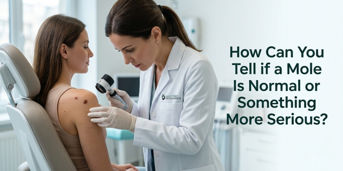

You catch a glimpse of a small brown spot on your forearm while applying sunscreen and pause. Has it always looked like that? Many people notice moles throughout life without issue, yet that moment of doubt is common. Sun exposure, genetics, and age all play roles in how these pigmented spots appear and evolve. The vast majority remain benign, but changes in size, shape, or color deserve prompt attention. Distinguishing normal variation from warning signs prevents unnecessary worry while ensuring timely care when needed. Expert evaluation at a Harley Street dermatology clinic provides clarity through thorough checks and advanced tools. This guide explains what makes a mole typical, which features raise concern, and how professional assessment supports informed decisions.
Early awareness of mole changes supports proactive skin care and timely professional guidance.
Understanding the Biology Behind Common Moles
Moles, also known as nevi, form when melanocytes — the cells that produce melanin pigment — cluster together instead of spreading evenly across the skin. This clustering often happens during childhood and adolescence due to genetic factors and sun exposure. Most moles are acquired over time, though some people are born with congenital ones. The pigment gives them their characteristic brown or tan color, though shades vary based on skin tone and depth. Normal moles stay stable in appearance, reflecting balanced cell activity. Changes can occur if cells behave irregularly, sometimes linked to cumulative UV damage or inherited traits. Recognizing this cellular foundation helps explain why monitoring matters. Not every variation signals trouble, but understanding the process empowers better self-observation and decisions about professional review.
Features of Typical, Harmless Moles
Typical moles share consistent traits that help differentiate them from concerning ones:
- Uniform Appearance: They usually display a single even shade of brown, tan, or pink, without patches of different colors. This consistency reflects stable melanocyte activity and is a reassuring sign during self-checks.
- Symmetrical Shape: Round or oval with smooth, well-defined borders that do not fade irregularly into surrounding skin. Symmetry indicates orderly growth patterns.
- Small Size: Most measure less than 6mm in diameter, roughly the width of a pencil eraser. Larger stable moles can exist but warrant occasional review.
- Stable Over Time: They do not change noticeably in size, shape, or color from month to month or year to year. Minor seasonal fading is possible but dramatic shifts are not typical.
- Smooth or Slightly Raised Texture: Surface feels even, sometimes with fine hairs, without crusting, scaling, or ulceration. Raised moles are often fleshier but maintain regular contours.
- No Associated Symptoms: They cause no itching, bleeding, tenderness, or inflammation unless irritated by clothing or shaving. Absence of discomfort supports a benign assessment.
- Common in Sun-Exposed Areas: They frequently appear on arms, face, back, and legs where UV exposure occurs, yet remain proportionate to overall skin pigmentation.
These characteristics provide a reliable baseline for personal monitoring between professional visits.
Warning Signs That Warrant Professional Attention
Moles that deviate from normal patterns deserve closer evaluation to rule out atypical changes or early melanoma.
The ABCDE Rule for Self-Assessment
This widely used guideline helps identify potential issues systematically. Asymmetry means one half does not match the other. Border irregularity shows notched, blurred, or scalloped edges instead of smooth outlines. Color variation includes multiple shades or unusual hues like red, white, or blue within the same mole. Diameter larger than 6mm raises attention, though smaller changing moles also matter. Evolving refers to any noticeable change in size, shape, color, or symptoms over time. Applying this rule regularly supports early detection without replacing professional judgment.
Additional Red Flags Beyond ABCDE
New moles appearing after age 40, bleeding without injury, persistent itching, or crusting can signal changes needing review. Moles on the scalp, soles, or palms require extra vigilance due to location. Rapid growth or elevation in previously flat moles also merits prompt check. These signs do not always indicate cancer but justify expert assessment for peace of mind.
An introductory line after this section helps frame next steps clearly. Consulting a specialist brings advanced tools like dermoscopy that reveal subsurface patterns invisible to the naked eye, offering greater accuracy than self-examination alone.
Watching for changes in color and shape is a simple yet vital habit for maintaining long-term skin health.
Common Risk Factors and When to Seek Evaluation
Several factors increase the likelihood of developing atypical moles or melanoma:
- Fair Skin and Sun History: Individuals with light skin, freckles, or history of sunburns face higher risk due to lower natural protection against UV damage.
- Family or Personal History: Previous melanoma, atypical moles, or close relatives with skin cancer call for more frequent checks and lower thresholds for removal.
- Large Number of Moles: People with over 50 moles have statistically elevated risk, making regular professional mapping valuable.
- Immune System Factors: Conditions or medications that suppress immunity can influence mole behavior and healing.
- Age and Hormonal Changes: New or changing moles in adulthood, especially during hormonal shifts, benefit from evaluation.
- Genetic Syndromes: Rare inherited conditions like dysplastic nevus syndrome warrant specialized monitoring from an early age.
Recognising these risks helps determine appropriate screening frequency and when mole removal treatment becomes a prudent choice for suspicious lesions.
Professional Diagnostic Techniques at Specialist Clinics
Dermatologists use precise methods beyond visual inspection to assess moles accurately. Dermoscopy provides magnified, illuminated views of pigment networks and vascular patterns, revealing details that distinguish benign from suspicious lesions. Digital mole mapping creates baseline images for ongoing comparison, ideal for patients with many moles. Biopsy techniques, such as shave or punch methods, allow laboratory analysis when visual clues are inconclusive. These approaches minimise unnecessary removals while ensuring concerning moles receive timely attention. At a Harley Street dermatology clinic, such tools combine with experienced clinical judgment for reliable outcomes. Patients receive clear explanations of findings and personalised recommendations, whether monitoring or proceeding to removal.
Treatment Options for Suspicious or Bothersome Moles
When evaluation indicates action, several approaches suit different situations:
- Surgical Excision: Complete removal with margins for suspicious moles, allowing full pathology review and low recurrence. Fine stitching supports excellent cosmetic results.
- Shave Removal: Suitable for raised, benign-appearing moles where deeper layers are not concerning. Quick healing with minimal scarring in many cases.
- Laser or Other Targeted Methods: Used selectively for certain pigmented lesions, though excision remains standard for potential melanoma risk.
- Cryotherapy: Occasionally applied to small benign lesions but less common for moles due to the need for tissue analysis.
- Combination Care: Post-removal scar management and sun protection advice help optimise long-term appearance.
- Preventive Strategies: Regular screenings and sun-safe habits reduce future risk alongside targeted removal of concerning moles.
Each option is discussed thoroughly so patients understand benefits, recovery, and expected outcomes.
Preparing for and Recovering from Mole Procedures
Proper preparation and aftercare contribute significantly to smooth experiences and optimal results.
What Happens During a Typical Consultation and Procedure
Clinicians review medical history, perform detailed examination, and discuss findings using visual aids when helpful. Local anaesthetic ensures comfort. The procedure is usually brief, with immediate wound care instructions provided. Many patients appreciate the calm, professional environment that reduces anxiety.
Post-Procedure Care and Healing Timeline
Keep the site clean and dry initially, following specific dressing guidance. Mild redness or swelling is normal and subsides quickly. Avoid strenuous activity or sun exposure on the area during early healing. Most stitches, if used, are removed within a week or two. Full maturation of the scar takes months but improves steadily with proper care. Patients receive guidance on signs that warrant follow-up contact.
These steps support safe healing and satisfying cosmetic outcomes while addressing individual concerns.
8. Common Myths and Misconceptions About Moles
Several widespread beliefs can lead to delayed care or risky self-treatment. Many assume all dark moles are dangerous, causing unnecessary alarm over stable, long-standing ones. Others believe moles with hair are automatically safe, overlooking that hair can grow from both typical and atypical lesions. The idea that home remedies or over-the-counter creams can safely remove or diagnose moles often results in irritation, incomplete treatment, or masking of important changes. Some think only sun-exposed moles matter, ignoring that melanoma can develop anywhere, including soles, palms, or under nails. Another myth suggests that once a mole stops growing it needs no further attention. In reality, any evolving feature justifies review. Dispelling these misconceptions encourages evidence-based decisions and timely professional input when features change.
Professional removal is the only way to ensure a growth is gone for good without risking permanent skin damage.
Modern Approaches to Mole Assessment and Management
Dermatology practices now integrate technology with clinical expertise for more precise care. Digital dermoscopy and total body photography create comprehensive records that track subtle shifts over years. Artificial intelligence-assisted tools can flag potential concerns for closer human review, though specialist interpretation remains essential. Patient education resources help individuals perform effective self-exams at home. Multidisciplinary teams coordinate when needed, linking dermatology with plastic surgery for optimal cosmetic results after removal. These advances support earlier detection and less invasive interventions when appropriate. Regular screenings tailored to personal risk profiles offer reassurance and proactive management. Combining professional tools with informed self-awareness creates a strong foundation for skin health.
- Technology Integration: High-resolution imaging and mapping systems allow detailed longitudinal tracking.
- Personalised Screening Plans: Risk assessment leads to custom intervals for full-body checks.
- Minimally Invasive Options: Refined techniques reduce scarring while maintaining thoroughness.
- Patient Support Resources: Educational materials and follow-up protocols enhance long-term adherence.
- Holistic Skin Health Focus: Advice on sun protection, lifestyle, and overall wellness complements mole-specific care.
Maintaining Healthy Skin and Ongoing Vigilance
Supporting skin health extends beyond individual mole checks. Daily broad-spectrum sunscreen use protects against UV-induced changes across all skin types. Protective clothing and shade-seeking habits during peak sun hours add meaningful layers of defence. Healthy lifestyle choices, including balanced nutrition and avoiding smoking, contribute to better tissue resilience. Self-examination routines performed monthly help familiarise you with your skin pattern so changes stand out. Scheduling periodic professional reviews, especially with personal or family risk factors, provides expert reassurance. After any mole removal treatment in London, continued sun safety and observation help preserve results and detect new developments early. These habits build confidence and contribute to lifelong skin wellbeing without constant worry.
Comparing Normal Moles with Suspicious or Atypical Ones
Feature | Normal Mole | Suspicious/Atypical Mole |
Symmetry | Evenly matched halves | Asymmetrical shape |
Border | Smooth, well-defined | Irregular, notched, or blurred |
Color | Uniform single shade | Multiple colors or uneven distribution |
Diameter | Usually under 6mm | Often larger, though size alone not decisive |
Evolution | Stable over time | Changing in size, shape, color, or symptoms |
Surface/Sensation | Smooth or evenly raised, no symptoms | Crusting, bleeding, itching, or ulceration |
FAQs About Moles and Skin Changes
How often should I check my moles at home?
Monthly self-exams in good lighting, using mirrors for hard-to-see areas, help track changes effectively between professional visits.
Can normal moles suddenly become cancerous?
While rare, changes in existing moles can occur. Any evolving features should prompt professional evaluation rather than waiting.
Does mole removal leave a noticeable scar?
Modern techniques at specialist clinics minimise scarring. Location, size, and individual healing influence final appearance, with good care improving outcomes.
Are all changing moles dangerous?
Not necessarily. Many changes are benign, but professional assessment determines whether monitoring or removal is appropriate.
What should I do if I notice a new mole as an adult?
New moles after age 30 or 40 benefit from dermatologist review to ensure they fit a normal pattern rather than atypical growth.
To Bring It All Together
Distinguishing normal moles from those requiring attention combines self-awareness with professional expertise. Typical moles show symmetry, uniformity, and stability, while changes in these features signal the need for evaluation. Tools like the ABCDE rule and advanced imaging support accurate assessment and timely mole removal treatment in London when indicated. Prioritising sun safety, regular checks, and specialist input builds confidence in managing skin health effectively. By staying informed and proactive, you support clear, healthy skin and peace of mind for years ahead.
Clarity on your skin’s condition leads to the confidence that you are providing it with the best possible care.